|
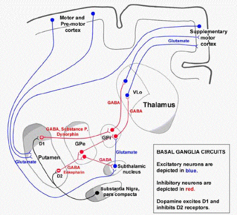
The role of the basal ganglia in voluntary movements is to initiate muscle activity and generate a smooth sequence of muscular actions that achieves the intended goal. The basal ganglia are at the centre of a series of neural circuits through which movements can be initiated and sequenced appropriate to events in our environment, memory, emotions or thought processes. The association cortex concerned with each of those functions is connected to a specific area of the basal ganglia, which in turn relays infomation back to the same area of the cortex via the thalamus. The premotor and supplementary motor cortices (Brodmann's area 6) and the bi-directional connections with the basal ganglia are essential for the planning, initiation, programming and sequencing of movements. The smooth sequencing of movements appears to depend on a balance between the activities of two neural pathways --direct and indirect - and the ability of two areas, the substantia nigra, and the subthalamic nucleus, to modulate this balance. Each area of association cortex has connections with a specific area of the basal ganglia, which in turn relays infomation back to the same area of the cortex via the thalamus. These networks appear to perform similar tasks irrespective of the function of the cortical area under consideration, such as visual/tactile/auditory maps of 3-dimensional space, attention, emotion, memory or voluntary movements. So subregions of the basal ganglia appear to be concerned with the functions of individual areas of association cortex. All of these areas cortical areas are also linked independently by subcortical association fibres. Afferent inputs from almost all areas of the cortex, appear to be processed within the basal ganglia using similar pathways consisting of (a) medium spiny neurones in the corpus striatum (b) neurones in the globus pallidus and (c) a relay station in the ventral thalamus, which (d) projects back to the area of cortex from which the afferents originated. A further neural looped circuit involving the substantia nigra and the modulatory effects of the subthalamic nucleus are linked into this network. The caudate nucleus, while anatomically distinct, behaves functionally as part of the striatum. One of the important connections of the basal ganglia is with Brodmann's area 6 which is involved in planning, organising and initiating voluntary movements, and includes the supplementary motor cortex (MII) and the pre-motor cortex. These areas project to the primary motor cortex (MI Brodmann's area 4) and become active before MI during voluntary movements. Together they are involved in organising sequences of movements into meaningful, goal-directed activity. Disorders such as Parkinson's Disease and Huntington's Chorea, in which tremors or uncontrollable involuntary movements are evident, are associated with degenerative changes within the basal ganglia. Below is a diagram of one of the parallel circuits linking an area of cortex with the basal ganglia and returning to the same area of cortex.  * *
GP= Globus Pallidus; Lateral GP= Globus Pallidus Externa; medial GP= Glodus Pallidus Interna Most cells in the corpus striatum are inhibitory neurones that release GABA at their nerve endings. They have complex dendritic fields densely covered with dendritic spines, hence the name "medium spiny neurons". Most of the synapses on these dendrites are excitatory, using glutamate as a neurotransmitter, and originate in the cortex. At rest, spiny neurons are hyperpolarised because they have a high potassium conductance, and consequently strong simultaneous excitatory inputs are necessary to fire them. Spiny neurones project to the globus pallidus, the medial part of the basal ganglia adjacent to the internal capsule, through which it connects with the thalamus. Neurones in the substantia nigra are also inhibited by the GABAergic spiny neurones, but both can become more active by the removal of the GABAergic influence; this removal of inhibition is known as disinhibition, and is the main mechanism by which the pallidal neurones become active.Within the sub-region of the basal ganglia associated with the premotor-cortex, these changes in the activity lead to movements, as a result of disinhibition within the striatum. There are also a number of other types of neurones in the striatum, by they account for only a few percent of the total. |
|
|
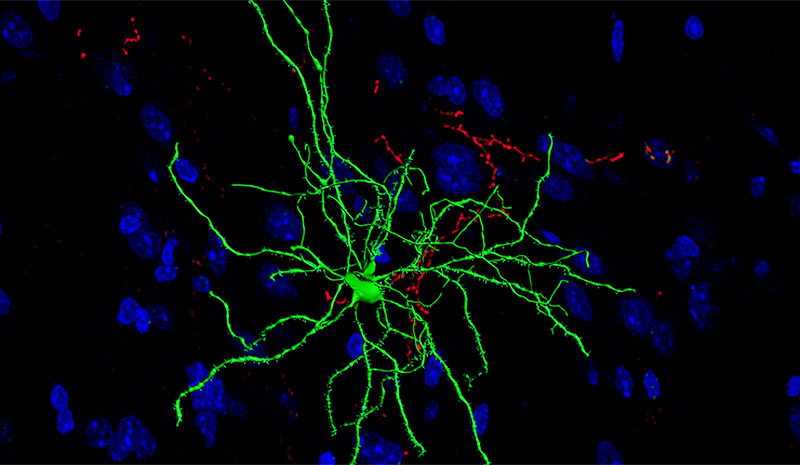
Medium spiny neurons are inhibitory interneurones that release GABA at their nerve endings, and form about 95% of the population of neurones in the striatum. The diagram above shows that dopamine, acting at different sets of synapses, has two opposing actions on medium spiny neurones, one being excitatory (D1) and the other inhibitory (D2). Note the dendritic spines on the medium spiny neurone (opposite). |
|
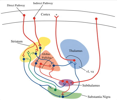
Direct and Indirect Pathways There appear to be two opposing pathways that process signals through the basal ganglia: one excites and the other inhibits the thalamic nuclei that provide feedback to the cortex. They are known as the direct pathway (which excites the thalamic neurones) and the indirect pathway (which inhibits them). In normal individuals there appears to be some sort of balance in this relationship. An imbalance in this relationship is thought to result in the motor changes seen in Parkinson's Disease. The direct pathway has been described above. Iit involves glutamergic afferents from the cortex exciting medium spiny neurones in the striatum; these inhibit GABAergic neurones in the Globus Pallidus interna, which in turn inhibit the thalamic neurones that return to the cortex. The indirect pathway has an additional relay in the subthalamic nucleus as shown opposite, which has excitatory effects on the GABAergic neurones that pass from the Globus Pallidus interna to the thalamus. NOTE; in the diagram opposite: Putamen=Corpus Striatum; GPe= Globus Pallidus Externa; GPi= Globus Pallidus Interna. |
|
|
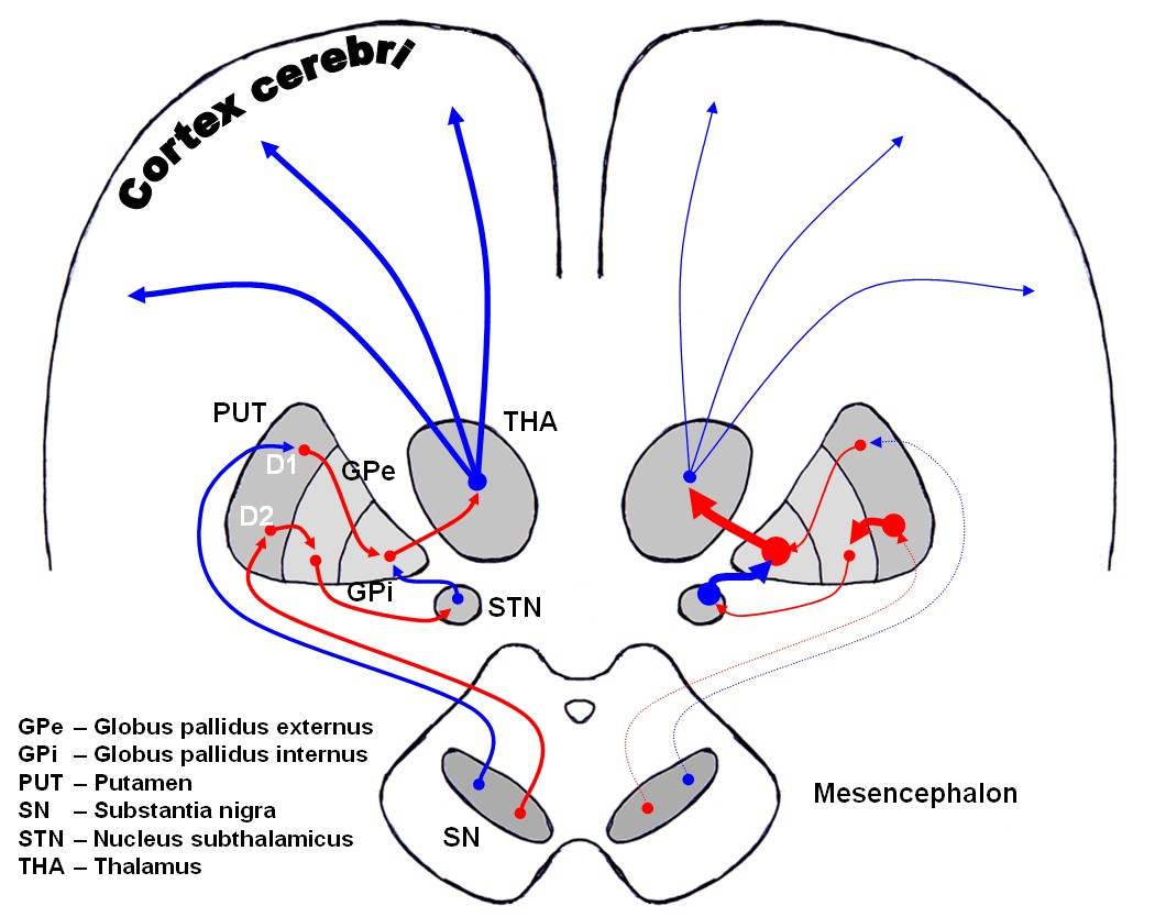
Substantia Nigra and the Nigro-Striatal Pathways The substantia nigra is located in the midbrain and helps to make movements occur in a smooth sequence; it is a dark thin plate on the dorsal surface of the cerebral peduncles, and its dark appearance is due to high levels of the pigment melanin in dopaminergic neurones that project to the basal ganglia in the nigro-striatal pathway. The Nigro-Striatal Pathways appear to be involved in modulating the direct and indirect pathways using dopamine as a transmitter. Striatal neurons on the direct pathway have D1 dopamine receptors, which depolarize the cell in response to dopamine. In contrast, indirect pathway striatal neurons have D2 dopamine receptors, which hyperpolarize the cell in response to dopamine. Because of these opposing actions, the nigrostriatal pathway can modulate the balance between these two pathways, exciting the direct pathway (using D1 receptors) while simultaneously inhibiting the indirect pathway (using D2 receptors). The nigro-striatal pathways degenerate in Parkinson's Disease, and is characterised by the presence of involuntary movements, such as a tremor at rest, and a poverty of movement and facial expression. This may be due to an alteration of the balance between the direct and indirect pathways. |
The nigro striatal pathways are shown on the left of this diagram. In Parkinson's disease, changes occur and the reduced size of the pathways and the consequences are shown on the right.
|
|
Direct and Indirect Pathways The diagram opposite shows the direct and indirect pathways and the connections of the subthalamic nucleus and substantia nigra. Deep Brain Stimulation Deep brain stimulation of the Subthalamic nucleus or other areas in the basal ganglia have been used in neurosurgical attempts to treat Parkinson's Disease. Deep brain stimulation is a surgical treatment involving the insertion of an electrode into specific areas of the basal ganglia, such as the subthalamic nucleus, so that electrical stimuli can be applied to those area. Deep brain stimulation has been an important advance in the treatment of Parkinson’s disease, and may help up to one fifth of patients . |
 * * |